Vetscan Imagyst product guides
AI Blood Smear Analysis
Tips
AI Blood Smear Analysis slide preparation
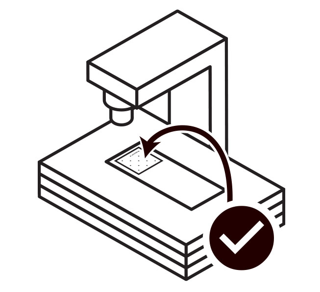
Scanning an AI Blood Smear Analysis slide
Reviewing AI Blood Smear Analysis results
AI Dermatology Diagnostics
Tips
AI Dermatology left and right ears sample preparation guide (PDF)
AI Dermatology infectious agent semi-quantitative category overview (PDF)
AI Dermatology Diagnostics slide preparation
Scanning an AI Dermatology Diagnostics slide
Reviewing AI Dermatology Diagnostics results
AI Equine Fecal Egg Count Analysis
AI Equine Fecal Egg Count (FEC) Analysis slide preparation
Reviewing AI Equine Fecal Egg Count (FEC) Analysis results
AI Fecal Analysis
Tips
AI Fecal Analysis slide preparation
AI Fecal Analysis coverslip placement
Scanning an AI Fecal Analysis slide
Reviewing AI Fecal Analysis results
AI Masses
AI Masses slide preparation
Adding an AI Masses test
Scanning an AI Masses slide
Reviewing AI Masses results
AI Urine Sediment Analysis
Tips
AI Urine Sediment complete urine sediment analysis point of care (PDF)
AI Urine Sediment hospital resource guide (PDF)
AI Urine Sediment medical whitepaper (PDF)
AI Urine Sediment Analysis dilution procedure (PDF)
AI Urine Sediment Analysis slide preparation
Scanning an AI Urine Sediment Analysis slide
Reviewing AI Urine Sediment Analysis results
Digital Cytology
Tips
Fluids
Body cavity (pericardial) (PDF)
Body cavity (pleural, peritoneal) (PDF)
CSF (cerebrospinal fluid) (PDF)
Synovial/joint (PDF)
Washes (PDF)
Digital Cytology sample preparation
Blood smear (PDF)
Digital Cytology: The basics (staining and submission) (PDF)
Improving suspected lipoma cytology samples (PDF)
Other sample types (PDF)
Tissue cytology (fine needle biopsy/aspiration) (PDF)
Digital Cytology slide template help
Adding a Digital Cytology test
Digital Cytology order entry
Digital Cytology slide preparation
Scanning a Digital Cytology slide
Fluid site card
Lesion site card
Reviewing Digital Cytology results
Quick Start Guides
AI Blood Smear Analysis Quick Start Guide (PDF)
AI Dermatology Diagnostics Quick Start Guide (PDF)
AI Equine Fecal Egg Count Analysis Quick Start Guide (PDF)
AI Fecal Analysis Quick Start Guide (PDF)
AI Masses Quick Start Guide (PDF)
AI Urine Sediment Analysis Quick Start Guide (PDF)
Digital Cytology Quick Start Guide (PDF)
Vetscan Imagyst user guide
Vetscan Imagyst overview
Company information
Gallery view
Adjust color
Assessment drawer
Carousel view
Gallery view
Gallery view actions panel
Gallery view features
Gallery view results panel
Images and objects
Help
My profile
Navigation
Order entry
Reports
Scanners
Grundium Ocus scanner overview
Grundium: locking and unlocking the slide lock
Grundium scanner troubleshooting guide
Routine scanner cleaning (PDF)
Scan area masks for Grundium scanners
Signing in, signing out, and session security
Signing in, signing out, and session security
Email (username)
Change password
Forgotten email (username)
Forgotten password
Slide view
Adding a field of view (FOV) box
Adding, editing, and deleting measurements
Editing an image capture
Exemplars gallery
Slide view
Slide view counts panel
Slide view features
Toggle measurements
Slide template help
Users
Workflow
Worklist
Video guides
AI Blood Smear Analysis
AI Blood Smear Analysis demonstration
AI Blood Smear Analysis do's and don'ts
AI Blood Smear enhanced workflow and reporting
AI Blood Smear Analysis using new methylene blue (NMB) stain
AI Dermatology Diagnostics
AI Dermatology demo
How to ear swab
How to impression smear
How to skin swab
How to submit a left and right ears test
AI Equine Fecal Egg Count Analysis
AI Fecal Analysis
Fecal check list
How to: run a fecal test
Fecal step 1: collect fecal matter
Fecal step 2: prepare the fecal sample
Fecal step 3: add the sample to a slide
Fecal step 4: place a coverslip on the slide
Fecal step 5: initiate the test
Fecal step 6: load and scan the slide
Fecal step 7: review the scan
AI Masses
Sample preparation: fine needle aspirate (FNA)
Sample preparation: fine needle biopsy (FNB)
How to run an AI Masses sample
AI Urine Sediment Analysis
AI Urine Sediment Add-on expert review with a stained slide
AI Urine Sediment demo
AI Urine Sediment sample preparation
Digital Cytology
Accessing Digital Cytology delivery options
Digital Cytology demo
How to: blood smear
How to: coverslip and immersion oil
How to: dos and dont’s
How to: ear cytology
How to: fine needle aspiration
How to: fine needle biopsy
How to: stain a slide
How to: urine sediment
How to: using the Imagyst application
Release notes
- All Categories
- Vetscan Imagyst product guides
- Digital Cytology
- Tips
- Digital Cytology slide template help
Digital Cytology slide template help

Errors can be reduced by following these simple steps
Single coverslip
Place only one coverslip on the slide.

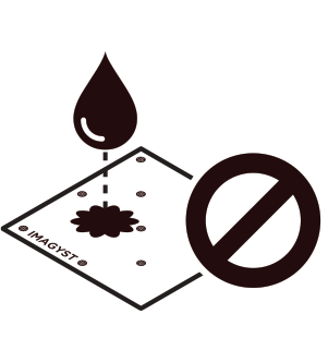
Dirty lens
Do not scan the slide if there is material on top of the coverslip as it will result in a dirty lens or scanning error.
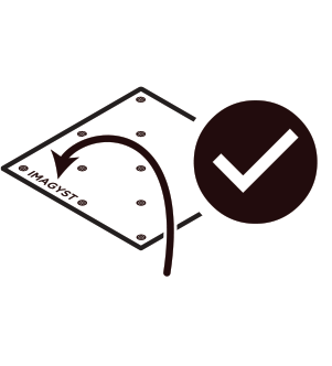
Imagyst placement
Place the coverslip so the Imagyst logo is in the location shown.
Slide placement
Ensure proper slide placement by placing the slide with the sample on the left.